From life-saving clinical diagnostics to advanced lab research – FLASH adapts to your workflow and elevates your results.


Innovation Meets Practical Application
Full-volume analysis
FLASH scans the entire tissue structure – not just surface slices – eliminating diagnostic blind spots and ensuring comprehensive, data-rich results.
Time & cost efficiency
Our streamlined workflows and automated processing reduce turnaround times and costs without compromising quality – making FLASH viable for real-world implementation.
Lab-ready kits
Pre-assembled, protocol-optimized kits designed for seamless integration into standard lab setups – with minimal hands-on time and maximum output.
Scalable impact
From diagnostics to discovery, FLASH is engineered to scale across clinical, academic, and pharmaceutical environments – without technical barriers.


Transforming Cancer Diagnostics with Full-Volume Precision
FLASH redefines lymph node analysis: comprehensive, cost-effective, and seamlessly integrable into clinical workflows. Detect more. Diagnose earlier. Improve outcomes.
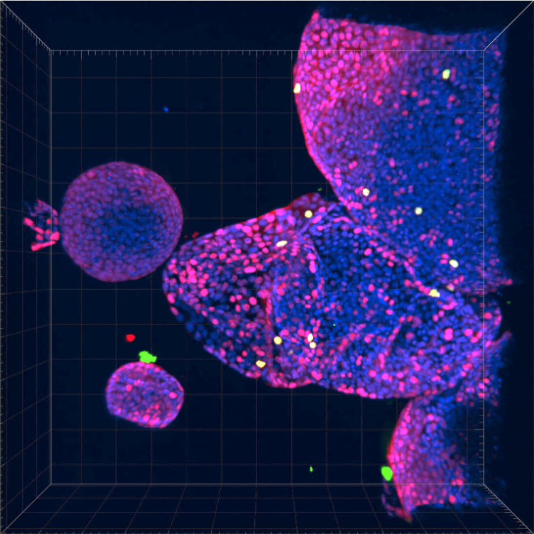
Scalable 3D Tissue Analysis Powered by FLASH Technology
Designed for sensitive and scalable 3D tissue culture analysis, the FLASH Organoid Imaging Kit brings the power of FLASH technology into research environments – from academic discovery to pharmaceutical screening ensuring reliable and reproducible results.
Our Expert Team and Esteemed Partners

Axel Behrens, PhD
Chief Scientific Officer (CSO)
Axel is a world-leading cancer researcher, director of the Cancer Research UK Convergence Science Centre, and a professor at both the Institute of Cancer Research and Imperial College London, with numerous prestigious accolades and memberships in Academia Europaea and EMBO.

Hendrik Messal, PhD
Chief Technology Officer (CTO)
Hendrik, first author of the groundbreaking Nature publication on FLASH technology and recipient of the 2022 Early Career Research Award by the Biochemical Society, is a leading cancer biologist known for his contributions to 3D whole organ imaging, cancer architecture, and 3D histopathology.

Karim Tabiti, PhD
Chief Executive Officer (CEO)
Karim is an experienced executive, having served as CEO of Biotype GmbH, led reagent and system business at Immunodiagnostic Systems, and held senior roles at Roche Diagnostics, specializing in product strategy, marketing, and commercialization.

Christine Munz, PhD
Advisory Board
Christine is a seasoned executive with 20+ years in life sciences and diagnostics, currently CEO at Eppendorf and formerly Vice President at Leica Biosystems, specializing in strategic growth and global operations.

Kurt Zatloukal, M.D.
Advisory Board
Kurt is a former Professor of Pathology at the Medical University of Graz and CEO of Zatloukal Innovations GmbH, is an expert in molecular pathology, biobanking, and biosafety, with over 284 publications and involvement in EU research programs and international standards.